成人网站亚洲二区乱码,少妇乳大丰满高潮喷水,久久久一本精品久久精品六六,国产精品白丝av网站在线观看,国产精品白丝av网站在线观看

24小時咨詢電話:400-991-0197

微博 微博|1(6)123.jpg 手機RTX123.jpg

產(chǎn)品中心
Home>產(chǎn)品中心>代理進口品牌>美國RD>Mouse TNF-alpha DuoSet ELISA

美國RD RD SYStems

Mouse TNF-alpha DuoSet ELISA

Mouse TNF-alpha DuoSet ELISA

美國RD

已獲點擊:

聯(lián)系方式

021-60348496

產(chǎn)品介紹
  • Assay Type
    Solid Phase Sandwich ELISA
  • Format
    96-well strip plate
  • Sample Type & Volume Required
    100 μL
  • Range
    31.20 - 2,000 pg/mL
  • Sufficient Materials
    For five or fifteen 96-well plates*
  • Specificity
    Please see the product datasheet
* Provided that the recommended microplates, buffers, diluents, substrates and solutions are used, and the assay is run as summarized in the Assay Procedure provided.
Ancillary Reagent Kit Available
DY008, DuoSet ELISA Ancillary Reagent Kit 2 - Please Inquire

This DuoSet ELISA Development kit contains the basic components required for the development of sandwich ELISAs to measure natural and recombinant mouse TNF-alpha. The suggested diluent is suitable for the analysis of most cell culture supernate samples. Diluents for complex matrices, such as serum and plasma, should be evaluated prior to use in this DuoSet.


Product Features
  • Optimized capture and detection antibody pairings with recommended concentrations save lengthy development time
  • Development protocols are provided to guide further assay optimization
  • Assay can be customized to your specific needs
  • Economical alternative to complete kits
Kit Content
  • Capture Antibody
  • Detection Antibody
  • Recombinant Standard
  • Streptavidin conjugated to horseradish-peroxidase (Streptavidin-HRP)

本文由專注于提供生物科技服務的齊一生物收集整理

Preparation and Storage
  • Storage
    Store the unopened product at 2 - 8 °C. Do not use past expiration date.
Background: TNF-alpha

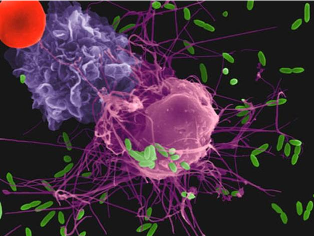
Tumor necrosis factor alpha (TNF-α), also known as cachectin and TNFSF2, is the prototypic ligand of the TNF superfamily. It is a pleiotropic molecule that plays a central role in inflammation, apoptosis, and immune system development. TNF-α is produced by a wide variety of immune and epithelial cell types. Human TNF-α consists of a 35 amino acid (aa) cytoplasmic domain, a 21 aa transmembrane segment, and a 177 aa extracellular domain (ECD). Within the ECD, human TNF-α shares 97% aa sequence identity with rhesus and 71% - 92% with bovine, canine, cotton rat, equine, feline, mouse, porcine, and rat TNF-α. The 26 kDa type 2 transmembrane protein is assembled intracellularly to form a noncovalently linked homotrimer. Ligation of this complex induces reverse signaling that promotes lymphocyte costimulation but diminishes monocyte responsiveness.

Cleavage of membrane bound TNF-α by TACE/ADAM17 releases a 55 kDa soluble trimeric form of TNF-α. TNF-α trimers bind the ubiquitous TNF RI and the hematopoietic cell-restricted TNF RII, both of which are also expressed as homotrimers. TNF-α regulates lymphoid tissue development through control of apoptosis. It also promotes inflammatory responses by inducing the activation of vascular endothelial cells and macrophages. TNF-α is a key cytokine in the development of several inflammatory disorders. It contributes to the development of type 2 diabetes through its effects on insulin resistance and fatty acid metabolism.

GENERAL ELISA PROTOCOL

Plate Preparation

  1. Dilute the Capture Antibody to the working concentration in PBS without carrier protein. Immediately coat a 96-well microplate with 100 μL per well of the diluted Capture Antibody. Seal the plate and incubate overnight at room temperature.
  2. Aspirate each well and wash with Wash Buffer, repeating the process two times for a total of three washes. Wash by filling each well with Wash Buffer (400 μL) using a squirt bottle, manifold dispenser, or autowasher. Complete removal of liquid at each step is essential for good performance. After the last wash, remove any remaining Wash Buffer by aspirating or by inverting the plate and blotting it against clean paper towels.
  3. Block plates by adding 300 μL Reagent Diluent to each well. Incubate at room temperature for a minimum of 1 hour.
  4. Repeat the aspiration/wash as in step 2. The plates are now ready for sample addition.

Assay Procedure

  1. Add 100 μL of sample or standards in Reagent Diluent, or an appropriate diluent, per well. Cover with an adhesive strip and incubate 2 hours at room temperature.
  2. Repeat the aspiration/wash as in step 2 of Plate Preparation.
  3. Add 100 μL of the Detection Antibody, diluted in Reagent Diluent, to each well. Cover with a new adhesive strip and incubate 2 hours at room temperature.
  4. Repeat the aspiration/wash as in step 2 of Plate Preparation.
  5. Add 100 μL of the working dilution of Streptavidin-HRP to each well. Cover the plate and incubate for 20 minutes at room temperature. Avoid placing the plate in direct light.
  6. Repeat the aspiration/wash as in step 2.
  7. Add 100 μL of Substrate Solution to each well. Incubate for 20 minutes at room temperature. Avoid placing the plate in direct light.
  8. Add 50 μL of Stop Solution to each well. Gently tap the plate to ensure thorough mixing.
  9. Determine the optical density of each well immediately, using a microplate reader set to 450 nm. If wavelength correction is available, set to 540 nm or 570 nm. If wavelength correction is not available, subtract readings at 540 nm or 570 nm from the readings at 450 nm. This subtraction will correct for optical imperfections in the plate. Readings made directly at 450 nm without correction may be higher and less accurate.

本文由專注于提供生物科技服務的齊一生物收集整理


返回

上一產(chǎn)品:Mouse TNF-alpha DuoSet ELISA

下一產(chǎn)品:Reagent Diluent Concentrate